Download
1 / 26
411 likes | 1.3k Views
ERYTHROPOIESIS. This takes place in the bone marrow and therefore, the first recognizable cell which can be identified as belonging to this series is the pronormoblast.

E N D
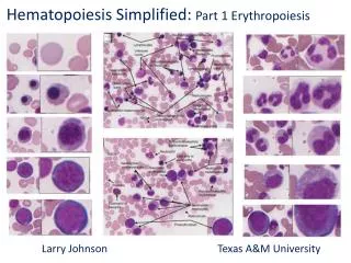
ERYTHROPOIESIS • This takes place in the bone marrow and therefore, the first recognizable cell which can be identified as belonging to this series is the pronormoblast. • It is from this cell that the red cells develop through a succession of maturing erythroblast namely basophilic/ early normoblast, polychromatic/ intermediate normoblast, orthochromatic/late normoblast.
Then from here it develops to reticulocyte and finally ends up with erythrocyte (mature red cell). • The whole process takes about 7 days. • This process of normoblastic maturation is characterized by the following progressive changes:-
1) The cell size diminishes. 2) Ripening of the cytoplasm. the staining reaction of the cytoplasm changes, the RNA starts diminishing) These two things take place simultaneously. a) So, colour changes to pink from blue due to reduction of RNA matter such that in Romanowsky stains,
there is a change in colour from deep blue to pink due to progressive formation of acidophilic staining haemoglobin and the simultaneous reduction of RNA which is responsible for basophilic of cytoplasm. b) Haemoglobin formation.
3) Ripening (maturing) of the nucleus having the large reddish, purple, open-network of the nucleus of a polychromatic normoblast converted to small deeply staining blue black structureless nucleus of the orthochromatic normoblast before it is eventually lost. i.e. the nucleus of the pronormoblast is larger than that of the orthochromatic normoblast.
The former (ponormoblast) has structures and stains reddish purple, while the later (orthochromatic) stains blue-black and is structureless. • Mitotic division of the developing cells in this series occurs up to the stage of the polychromatic normoblast and therefore the orthochromatic normoblast is not considered capable of mitotic division.
Characteristics of developing normoblast in romanowsky stained film. 1. Pronormoblast • Pronormoblast is a large oval cell. It comes about as a result of activation by erythropoeitin (hormone) which is produced in the kidney.
Cont: • This hormone acts on erythroid responsive cell. • The cytoplasm is more and stains deep blue. • The nucleus is round and occupies most of the cell and has one or two nucleoli. • The cell measures 12-20 micrometre in diameter.
2. Basophillic normoblast : (Early) • The size varies from 10-16 micrometer. • The nucleus is relatively large but smaller than pronormoblast stage. • The cytoplasm is smaller to that of pronormoblastic but slightly stage (plentiful).
The chromatic strands are more thicker and more deeply staining living a coarser appearance due to RNA. • At this stage the nucleus have disappeared.
3. Polychromatic normoblast (intermediate) • They vary from 8/14 micrometer in diameter. • The nucleus is smaller and occupies a smaller part of the cell. • It is reddish purple in colour. • The cytoplasm is large and begins to acquire haemoglobin, thus taking acidophillic – pink or pale pink colour.
4. Orthochromatic normoblast (late) • The size ranges from 8-10 micrometer in diameter. • The nucleus is small with condensed homogenous structereless chromatin. • It stains blue-black colour. The nucleus is eccentric and sometimes lobulated.
The cytoplasm is typically acidophillic (pink) because haemoglobinization has started taking place which gives it acidophillic staining. • The nucleus is lost at this stage.
5. Reticulocytes • It is a flat disc shaped and non-nucleated cell larger than a mature red blood cell. • When stained with romanowsky stains shows the diffuse oale basophillia. • The cell is called polymetachromatophillic cell, while with supra vital stains, the basophillic material appears in the form of reticulum and chromatic strands, which stains dark red.
Cont: • The Hb content is almost the same as that of mature RBC. • From the stage of retic to mature RBC takes about 1-2 days. • The nucleus is injected out at the spleen and the RNA and any other inclusions are removed and then the cell is released in to the circulation in the blood stream.
As the cell matures it becomes smaller in size and the nucleus diminishes and eventually lost i.e. the chromatin becomes less dense and the nucleus diminishes and eventually lost. • Nucleoli disappear before the nucleus is lost. • At this stage the cytoplasm losses the protein synthesis property and acquires haemoglobin.
6. Erythrocyte • The nucleus is not present. • The biconcave orange cytoplasm has the paler center occupying a third of the cell area. • The size is about 6.7-7.7 micrometer in diameter and 2.5 micrometer thick. • The outline is regular.
Characteristics of erythrocytes • When unstained, they appear as non-nucleated pale greenish yellow biconcave discs. • When stained by the romanowsky stain, they have affinity for eosin and therefore they stain pinkish under a microscopic exam. • Their normal range,
Women – 3.9-5.9m/mm3 Men- 4.5-6.5 m/mm3 Children – 4-6 m/mm3 • Their life span is 110-120 days after which they are removed by the R.E.S where they are broken down to some of their constituents, some of which are reutilized e.g. Fe and protein for haemopoiesis.
When placed in hypotonic saline, they swell and loose the biconcavity and assume a different shape (spherical). • They contain the red pigment called haemoglobin. • They have no granules. They are living cells. They contain enzymes of both anaerobic glycolytic pathway of (pentose, phosphate pathway).
60% of RBC is H2O and 40% is solid dry substance of which 90% is haemoglobin and 10% is stroma. • RBC are flexible thus they can readily be destroyed or distorted in shape e.g. when passing through small capillaries but quickly resume their normal shape after passing.
Cont: • Outside the body they behave abnormally like they tend to clump together a condition known as rouleaux formation. • When the fluid is withdrawn out of the cell membrane (hypertonic solution) the surface tends to shrink and form a number of knobs or lobes of irregular shape. • This is called crenation. • The biconcave shape gives the red cell the affinity to O2
Functions 1. It transports oxygen from the lungs through the heart to the tissues and carbon dioxide from the tissues to the lungs and finally out of the body. 2. They carry antigens. 3. They contain haemoglobin.
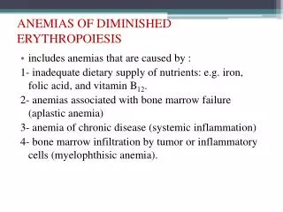
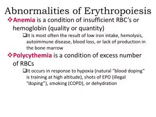
Cont: Types of erythropoiesis 1. Microcytic erythropoiesis • This is where cells are produced being smaller in size. It happens because of disease conditions e.g. iron deficiency anaemia, thalassaemia etc.
2. Macrocytic erythropoiesis • Cells are produced being larger in size due to abnormal conditions e.g. vitamin B12 / Folic acid deficiency. Also in haemolytic anaemia. 3. Normocytic / normoblastic erythropoiesis. • It gives rise to normal red blood cells.
Requirement of erythropoiesis • This can be divided into 2 categories: a) nutritional requirement. b) non nutritional requirement. • Nutritional requirements deal with diet e.g. iron, vitamin, folates, trace metal, amino acids. • Non nutritional requirements include hormones e.g. adrenaline, erythropoietin steroids.