
What are reticulocytes and why are they important in pathology?
Reticulocytes are immature red blood cells that still contain remnants of ribosomal RNA. They are important in pathology because they serve as indicators of bone marrow activity and can provide valuable information about various hematological disorders.
How are reticulocytes formed and what are its functions?
They are formed from hematopoietic progenitor stem cells through the process called erythropoiesis. Click on the link to read more on erythropoiesis https://ilovepathology.com/erythropoiesis/
Functions of reticulocytes
a. Hemoglobin Synthesis: Reticulocytes play a crucial role in the production of hemoglobin, the oxygen-carrying molecule in red blood cells. As reticulocytes mature into erythrocytes, they accumulate hemoglobin, which enables them to effectively transport oxygen from the lungs to various tissues and organs in the body.
b. Indicator of Bone Marrow Activity: The presence and quantity of reticulocytes in the bloodstream serve as an indicator of bone marrow activity. An increased reticulocyte count suggests active erythropoiesis and can be a compensatory response to conditions such as anemia or blood loss. Conversely, a decreased reticulocyte count may indicate reduced or impaired bone marrow function.
c. As diagnostic marker: reticulocyte counts along with indices are valuable in the evaluation of various hematological disorders, helps to differentiate different types of anemia and thus help in proper management of these diseases.
How are reticulocytes different from mature red blood cells?
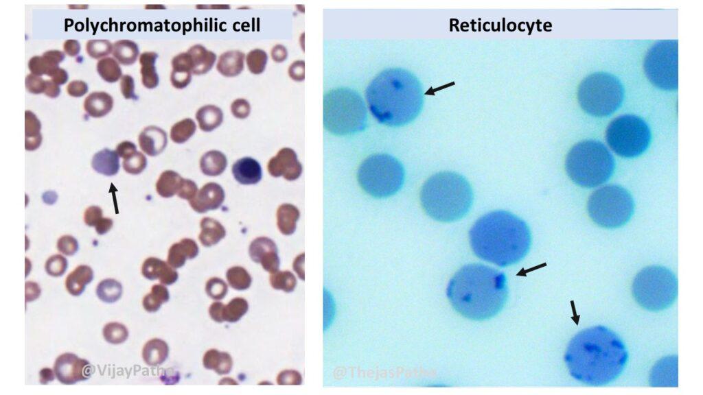
Unlike mature red blood cells, reticulocytes still have remnants of RNA and organelles making them larger. Ribosomal RNA, gives them a bluish hue when stained with certain dyes, such as Wright’s stain.
This blue color is often a mixture of the blue stain and the pink-red color of the hemoglobin within the reticulocytes. Hence these cells appear as a combination of blue and pink or reddish hues, hence the term “polychromatophilic” (poly = many, chroma = color).
Reticulocytes also have a shorter lifespan compared to mature red blood cells. On average, reticulocytes circulate in the bloodstream for about 1 to 2 days before they fully mature into erythrocytes.
How are reticulocytes demonstrated ?
Reticulocytes can be demonstrated and visualized through various laboratory techniques. Some common methods include
a. Supravital Staining:
b. Automated Hematology Analyzers
c. Fluorescent Staining:
a. Supravital staining: The term “supravital stain” is used to describe a staining method that is performed on living cells or tissues.
The term “supravital” itself originates from Latin, where “supra” means “above” or “beyond,” and “vitalis” means “life.” In the context of reticulocytes, the red blood cells are stained while they are still viable, without the need of fixation.
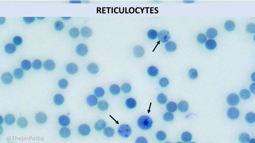
The reason behind using supravital staining for reticulocytes is to preserve their characteristic features, particularly the remnants of ribosomal RNA, which give them their RETICULAR appearance. And hence the term “reticulocyte” for these polychromatophilic cells (on Leishman/wright stain)
b. Automated Hematology Analyzers: These use various principles, such as flow cytometry or impedance-based methods, to identify and quantify reticulocytes in a blood sample.
c. Fluorescent Staining: Fluorescent staining methods, such as thiazole orange or acridine orange staining, can be used to identify and quantify reticulocytes. These stains bind to the RNA in reticulocytes and emit fluorescence when excited by specific wavelengths of light. Flow cytometry or fluorescence microscopy is then used to detect and analyze the stained reticulocytes.
What are the normal reference ranges?
Newborn : 2.5 to 6.5%
Infant: 0.5 to 3.1%
child/Adults/elderly : 0.5 to 2.5%
What is absolute reticulocyte count?
The absolute reticulocyte count is a measure of the actual number of reticulocytes present in a specific volume of blood.
The absolute reticulocyte count is typically reported as the number of reticulocytes per microliter (µL) or liter (L) of blood. It is calculated by the following formula:
Absolute Reticulocyte Count (per µL) = Reticulocyte Percentage × Red Blood Cell Count (in millions/µL) / 100
What are the causes of increased reticulocyte counts?
The reticulocyte count is increased in
a. Hemolytic anemias
b. Acute or chronic blood loss
c. Response to treatment of anemias
d. Increased production of erythropoietin- as response to hypoxia. Eg-high altitude, chronic lung disease
What are the causes of decreased reticulocyte counts?
This is also called as reticulocytopenia. The reticulocyte counts are decreased in
a. Bone marrow disorders: Aplastic anemia, myelodysplastic syndromes and leukemia
b. Nutritional anemias: Iron and B12/folate deficiency anemias
c. Infiltrative diseases of Bone marrow: Lymphoma, myeloma or metastasis
d. Infections : Parvovirus B infection, HIV etc
e. Chronic infections/inflammations : due to suppression of erythropoiesis or decrease in erythropoietin
f. Chemoradiation resulting in suppression of erythropoiesis.
What is Corrected reticulocyte count?
This is calculated by using the formula
[%reticulocyte count x Patient Hct]/45(normal Hct)
This is an anemia-specific corrected reticulocyte count.
A reticulocyte count that is artificially raised can happen in anemias. Even though erythropoiesis has not yet occurred, the reticulocyte count automatically rises when RBCs decline because it is a percentage of RBCs. It’s possible that the adequate increase in erythropoiesis in response to the anemia has not happened. Therefore, this value corrects for this problem.
What is immature reticulocyte fraction?
The Immature Reticulocyte Fraction (IRF) is a measure of the percentage of reticulocytes that are considered “immature” or recently released from the bone marrow. It provides information about the proportion of reticulocytes that are newly formed and still in the maturation process.
The IRF calculation is as follows:
IRF = Immature Reticulocyte Count / Total Reticulocyte Count
What are the differences between mature and immature reticulocytes
| Aspect | Mature Reticulocytes | Immature Reticulocytes |
|---|---|---|
| Appearance | Slightly larger size and faint bluish tint | Slightly smaller size and bluish coloration |
| RNA Content | Minimal to no visible remnants of RNA | Visible remnants of RNA |
| Nuclear Features | No visible nucleus | Residual nuclear material or remnants of the nucleus |
| Staining Pattern | Fine reticular or granular appearance | More intense and coarse staining pattern |
| Development Stage | Final stage before becoming mature erythrocytes | Recently released from the bone marrow and still undergoing maturation |
| Functional Status | Close to fully functional red blood cells | Still in the process of maturation and acquiring functional characteristics |
| Duration in Bloodstream | Shorter lifespan, soon to become fully mature erythrocytes | Recently released and transient presence in the bloodstream |
| Role | Oxygen transport and gas exchange | Preparation for becoming fully mature erythrocytes and contributing to oxygen transport |
| Significance | Indicates a stage close to mature erythrocytes | Indicates ongoing erythropoiesis and bone marrow activity |
What is Reticulocyte Hemoglobin Content (Chr) ?
Reticulocyte Hemoglobin Content (Chr) is a parameter that measures the amount of hemoglobin within reticulocytes. It provides information about the hemoglobinization or iron content of reticulocytes, reflecting their ability to synthesize hemoglobin during the maturation process.
Chr is reported as a numerical value or as a mean corpuscular hemoglobin concentration (MCHC) equivalent.
It helps evaluate the efficiency of hemoglobin synthesis and can be used as a parameter for assessing iron deficiency or iron-restricted erythropoiesis.


Recent Comments